Orlando Diagnostic Center
Imaging Services
Six specialized diagnostic modalities — each performed by experienced technologists and read by board-certified radiologists at our ACR-accredited centers.
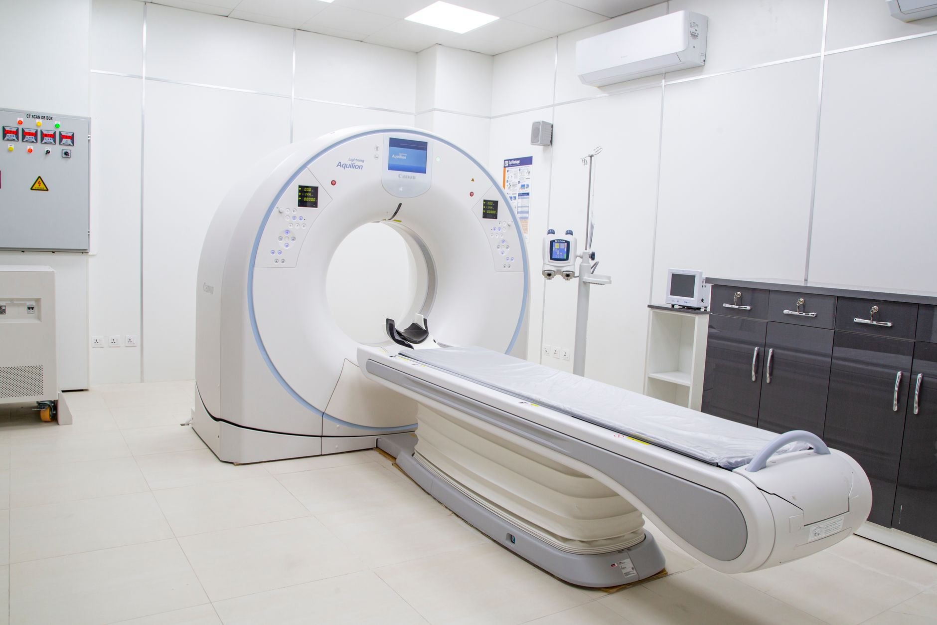
Magnetic Resonance Imaging (MRI)
Detailed soft-tissue imaging without radiation.
MRI uses a powerful magnetic field and radio waves to produce detailed, high-resolution cross-sectional images of internal structures. It is particularly valuable for evaluating the brain, spine, joints, and soft tissues throughout the body.
Our ACR-accredited MRI suite supports a wide range of clinical indications, including neurological evaluation, musculoskeletal assessment, abdominal and pelvic imaging, and vascular studies. Because MRI does not use ionizing radiation, it is often the preferred modality for repeated studies and for patients who require comprehensive soft-tissue detail.
Your MRI study is read by a board-certified radiologist on staff, and the signed report is delivered to your referring physician typically within 24 to 48 hours.
Preparation
Some MRI exams require preparation, such as fasting or a contrast injection. Your referring physician's order will specify any requirements, and our scheduling team will walk you through what to expect before your appointment.
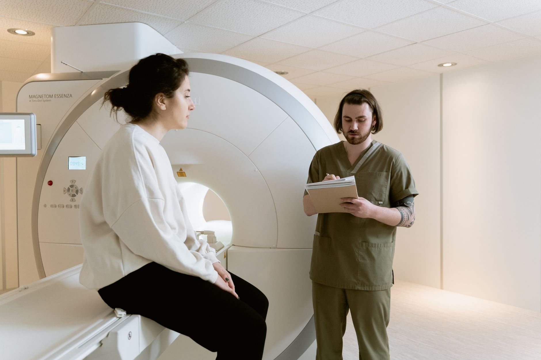
CT Scan (Computed Tomography)
Fast, precise cross-sectional imaging for complex diagnoses.
CT scanning combines a series of X-ray images taken from different angles and uses computer processing to create detailed cross-sectional images of bones, blood vessels, and soft tissues inside the body. It is faster than MRI and particularly effective for evaluating trauma, pulmonary conditions, abdominal concerns, and vascular structures.
Our CT protocols are optimized for low-dose radiation while maintaining the diagnostic image quality your physician requires. Both our Longwood and Kissimmee centers are equipped with modern multi-slice CT systems capable of handling a full range of diagnostic indications.
Results are read by a board-certified radiologist and delivered to your referring physician promptly — often the same day for routine studies.
Preparation
Depending on the study, you may be asked to fast for a few hours before your CT appointment, particularly if intravenous contrast is ordered. Our scheduling team will provide specific instructions when you book.
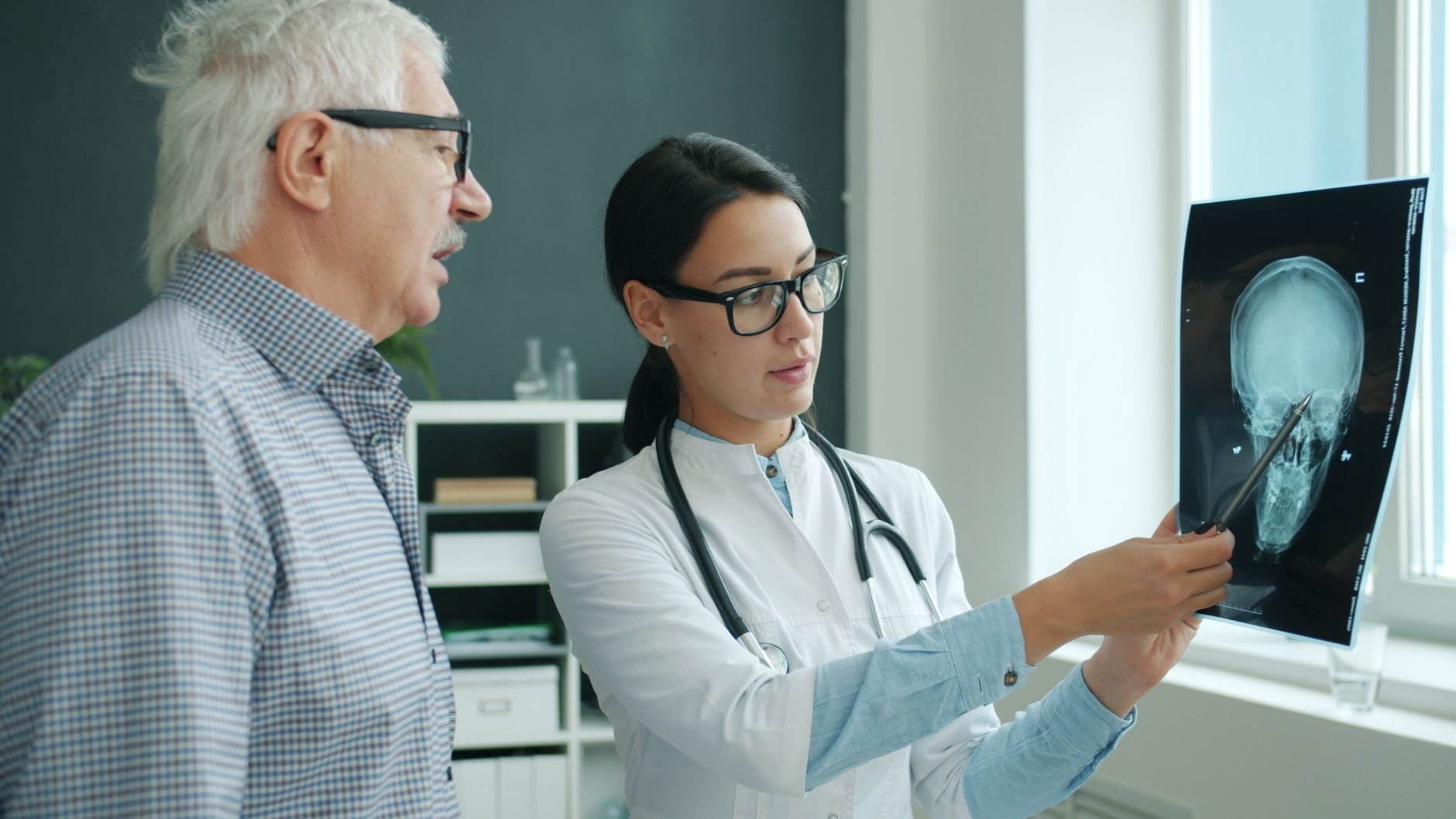
Echocardiogram
Real-time cardiac ultrasound for precise heart assessment.
An echocardiogram is a non-invasive ultrasound study of the heart. Using high-frequency sound waves, it generates real-time images of the heart's chambers, valves, walls, and great vessels, allowing your cardiologist or physician to assess cardiac structure and function in detail.
We perform transthoracic echocardiograms (TTE) — the most common type — which evaluate ejection fraction, wall motion, valve function, and pericardial conditions. The study is safe, painless, and carries no radiation exposure.
Results are interpreted by our board-certified radiologists and returned to your referring cardiologist or physician, typically within 24 to 48 hours.
Preparation
No special preparation is required for a standard transthoracic echocardiogram. Wear comfortable, loose-fitting clothing to your appointment. The study takes approximately 45 to 60 minutes.
Digital X-Ray
Immediate, high-resolution images with minimal radiation.
Digital X-ray (radiography) produces immediate, high-resolution images of bones and certain soft tissues. It is the first-line imaging study for evaluating fractures, joint conditions, chest pathology, and many orthopedic concerns.
Our digital systems provide superior image quality compared to older film-screen technology, while delivering less radiation to the patient. Images are acquired almost instantly, and reports are typically completed the same day. Digital X-ray studies serve as an efficient entry point that often guides the need for additional cross-sectional imaging.
X-ray studies are read and signed by a board-certified radiologist on staff.
Preparation
No preparation is required for most X-ray studies. You may be asked to remove jewelry or clothing with metal fasteners for the region being imaged. Studies are completed in minutes.
Ultrasound
Sound-wave imaging — safe, fast, and radiation-free.
Ultrasound uses high-frequency sound waves to produce real-time images of internal organs, blood vessels, and soft tissues. It is safe for all patient populations, carries no radiation exposure, and is often the imaging modality of choice for evaluating abdominal organs, the gallbladder, kidneys, pelvis, thyroid, and vascular structures.
We offer a broad range of ultrasound examinations, including abdominal, pelvic, vascular (Doppler), and musculoskeletal studies. Real-time imaging allows the technologist to evaluate structures dynamically, and the images are then interpreted and reported by our board-certified radiologists.
Ultrasound is also the primary imaging tool during pregnancy and for guiding certain diagnostic procedures.
Preparation
Preparation depends on the study. Abdominal ultrasound typically requires fasting for 4 to 6 hours. Pelvic ultrasound may require a full bladder. Our scheduling team will provide the specific preparation instructions for your study when you book.
DEXA / Bone Density Scan
The clinical standard for measuring and monitoring bone health.
DEXA (Dual-Energy X-ray Absorptiometry) is the most accurate and widely used test for measuring bone mineral density (BMD). It is essential for diagnosing osteoporosis, assessing fracture risk, and tracking the response to osteoporosis treatment over time.
DEXA uses two low-dose X-ray beams to measure the density of bone — typically the lumbar spine and hip — and compares the result to established reference norms. The radiation dose is very low, making it safe for routine monitoring.
Results include a T-score and Z-score, which your physician uses to classify bone density status (normal, osteopenia, or osteoporosis) and guide treatment decisions. Scans are read and reported by our board-certified radiologists.
Preparation
No fasting is required. Avoid calcium supplements for 24 hours before the exam. Wear comfortable clothing without metal zippers or buttons. The scan takes approximately 20 to 30 minutes.
FAQ
Questions About Our Services
Answers to what patients and referring physicians most commonly ask about our imaging services.
What imaging services does Orlando Diagnostic Center offer?
We offer six modalities: MRI, CT Scan, Echocardiogram, Digital X-Ray, Ultrasound, and DEXA/Bone Density scanning. All services are performed at both our Longwood and Kissimmee locations and are read by board-certified radiologists on staff.
Do I need a doctor's order to get imaging?
Most imaging studies require a physician's order. Your doctor's office can send the order directly to us by fax or electronic referral. Once we receive it, our scheduling team will contact you to confirm your appointment and provide any preparation instructions.
How do I know which imaging service I need?
Your referring physician determines the appropriate imaging modality based on your clinical situation. If you have questions about which study has been ordered, your doctor's office is the best resource. Our scheduling team is also happy to answer general questions.
Are contrast agents or dyes used in MRI or CT?
Some MRI and CT studies use intravenous contrast to enhance visibility of certain tissues and blood vessels. Whether contrast is required depends on the specific study ordered by your physician. If contrast is needed, our team will screen you for relevant allergies and kidney function prior to the exam.
How are my results delivered to my doctor?
Your study is interpreted by a board-certified radiologist who generates a signed radiology report. That report is delivered electronically to your referring physician, typically within 24 to 48 hours for routine studies. Urgent findings are communicated more rapidly.
Have another question? Contact our scheduling team — we're glad to help.
Ready to Schedule?
Request an appointment online and our scheduling team will confirm the details and any preparation instructions by phone.
Request an AppointmentNo referral required for most services. We accept most major insurance plans.